ads

Ads

  • Home
  • Eye Health
  • Men Health
  • Women Health

Ads2

Health & Wellness

If you buy something through a link on this page, we may earn a small commission. How this works.

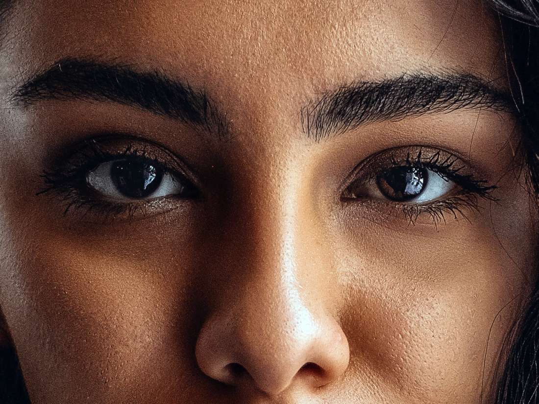
Asymmetrical eyes — or eyes that are not the same size, shape, or level as each other — are very common.

In rare cases, having asymmetrical eyes may indicate an underlying medical condition. Most of the time, however, this is not a cause for concern.

Although a person may be aware of their own facial asymmetry, it is unlikely that others will notice.

In fact, most people have asymmetrical features, with research indicating that some degree of facial asymmetry is both normal and desirable.

Read on to learn more about asymmetrical eyes, including some potential causes and home remedies.

Potential causes of asymmetrical eyes include:

Genetics

Genetics can account for uneven eyes and other types of facial asymmetry.

People with asymmetrical eyes may notice that other members of their family have similar features.

Having asymmetrical eyes as a result of genetics is not a cause for concern.
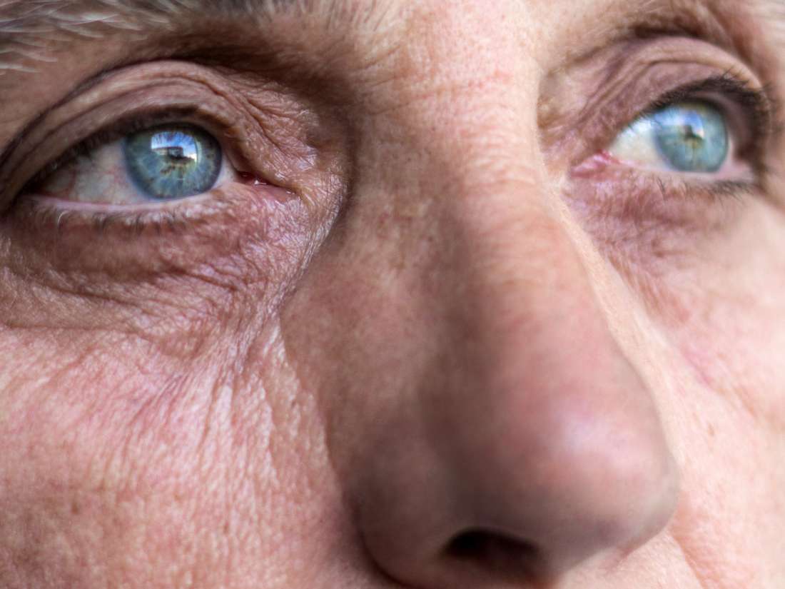
Aging

Imaging studies show a significant link between increasing age and facial asymmetry.

As people age, the soft tissues in the face relax. Cartilage, such as that in the nose, continues to grow while the bones do not. These changes can cause asymmetry.

Lifestyle factors

Some lifestyle factors can contribute to uneven eyes. For example, research on sets of twins has linked smoking with upper eyelid ptosis, also known as droopy eyelids.

Also, excessive sun exposure can change the skin around the eyes. Sun damage may affect one side of the face more than the other, leading to asymmetry.

Bell's palsy

Bell's palsy is a type of sudden, temporary facial paralysis. It causes one side of the face to droop, affecting the smile and one eye.

Its cause is currently unknown, though it may be due to trauma, nerve damage, or a complication of a viral infection.

Other signs and symptoms of Bell's palsy include:

  • changes in tear or saliva production
  • difficulty making facial expressions
  • drooling
  • headaches
  • jaw or ear pain

Trauma

Sustaining a blow to the face or being involved in a vehicle collision can cause damage to the eye area, leading to asymmetry.

Facial trauma may cause enophthalmos, or displacement of the eye. This causes people to appear as if they have a sunken eye.

Sinus conditions

Some sinus conditions can also lead to enophthalmos. These include:

  • chronic maxillary sinusitis
  • maxillary sinus tumors
  • silent sinus syndrome

With these conditions, changes to the eye can happen suddenly or gradually. They may also cause other symptoms, including:

Graves' disease

Graves' disease is an autoimmune condition that causes an overactive thyroid (hyperthyroidism).

People with Graves' disease can develop proptosis, or bulging eyes. When this affects one eye more than the other, it can lead to asymmetry.

Some other signs and symptoms of Graves' disease include:

  • anxiety
  • changes in sexual desire or function
  • enlargement of the thyroid gland (goiter)
  • fatigue
  • heart palpitations
  • menstrual changes
  • sensitivity to heat
  • sweating
  • unintended weight loss

Stroke

Stroke is a medical emergency. It can occur when there is reduced blood flow to the brain.

People can develop sudden facial asymmetry due to stroke. If the drooping is extreme, it may affect a person's vision.

Other symptoms of stroke include:

  • difficulty speaking and understanding
  • a sudden, severe headache
  • loss of balance or coordination
  • numbness or weakness of the face, one arm, and one leg
  • sudden onset of blurred or double vision

In most cases, uneven eyes do not require treatment. This is especially true if the asymmetry is the result of genetics or aging.

However, if an underlying medical condition is contributing to facial asymmetry, people may require treatment for the condition. Treatment may also be necessary if asymmetrical eyes are causing vision problems.

Some people may wish to treat uneven eyes for cosmetic reasons.

Possible treatments include:

Addressing underlying medical conditions

In some cases, treating the underlying medical condition responsible can make asymmetrical eyes seem less noticeable.

For example, treating Graves' disease with radioactive iodine or thyroid medications may stop the eyes protruding.

Those who have a medical condition that is contributing to their eye asymmetry should speak to their doctor about managing their symptoms.

Botox

Botox is a nonsurgical option for facial asymmetry. It involves injecting Botox, which is a muscle relaxer that comes from the bacterium Clostridium botulinum, into the area around the eyebrows.

Botox treatment lifts the brows, reducing the appearance of uneven eyes. The effects of Botox will typically last for around 3–6 months.

Brow lift

A brow lift is a cosmetic procedure that elevates the eyebrows. The aim is to give the face a more youthful appearance and provide greater facial symmetry.

There are different types of techniques a surgeon might use to lift the brow, but they will usually perform the procedure while a person is under general anesthesia.

Some potential risks of a brow lift include:

  • bleeding
  • further asymmetry (though additional surgery can correct this)
  • hair loss or changes to the hairline
  • infections
  • an allergic reaction to the anesthetic
  • scarring
  • temporary or permanent skin numbness

The results of a brow lift are not permanent. Aging and sun damage can cause the skin to droop again.

Blepharoplasty

Blepharoplasty is a type of cosmetic surgery that corrects uneven eyelids. It is a frequently performed aesthetic procedure.

During the procedure, a surgeon will remove excess fat, muscle, or skin from around the eye area to make the eyes appear more symmetrical.

After this surgery, a person may experience temporary bruising and swelling.

Some other risks include:

  • bleeding
  • infections
  • an allergic reaction to the anesthetic
  • scarring

Less commonly, the procedure may cause chronic conjunctivitis (inflammation of a part of the eye) or swelling that lasts for more than 3 months.

In rare cases, blindness can occur.

Orbital surgery

Orbital surgery is surgery on the eye socket (orbit). There are a few different types of orbital surgery, depending on the problem and the area of the eye socket that it affects.

A surgeon may carry out procedures to:

  • repair fractures
  • remove tumors
  • remove bones or fat to treat the effects of Graves' disease
  • reconstruct the anatomy of the socket

Like all surgeries, these procedures carry risks.

If they wish to, people with minor facial asymmetry may be able to use home remedies to make their eyes appear more symmetrical.

Some options include:

Makeup techniques

Various contouring and highlighting techniques can reduce uneven eyes and eyebrows. Makeup artists and online tutorials can provide guidance on this.

Some people even use hairstyling techniques to draw attention away from their eyes.

Eyelid tape

Putting eyelid tape on a sagging eyelid can lift the skin, hiding the sagging and asymmetry.

These thin, transparent strips are available to buy in beauty stores. They are also available online.

Asymmetrical facial features are normal and common. They are often the result of genetics, aging, or lifestyle factors.

Most people do not notice facial asymmetry in others, and research shows that it may even be a desirable feature.

However, for those who wish to address uneven eyes, several cosmetic procedures and home remedies are available.

In some cases, a medical condition may be causing facial asymmetry. In these cases, treating the underlying condition may help reduce the appearance of uneven eyes.

Anyone concerned about their facial asymmetry can speak to their doctor. It is also a good idea to seek medical attention if the asymmetry came on suddenly or if it is causing vision problems or other symptoms.

We picked linked items based on the quality of products, and list the pros and cons of each to help you determine which will work best for you. We partner with some of the companies that sell these products, which means Healthline UK and our partners may receive a portion of revenues if you make a purchase using a link(s) above.


Read More

An analysis of how the retina of the eye scatters light shows promise as an aid for the early diagnosis of Alzheimer's disease.

close up of an eyeShare on Pinterest
Examining how the retina disperses light can provide insight into Alzheimer's disease.

Scientists from the University of Minnesota in Minneapolis came to this conclusion after carrying out a recent study, the findings of which appear in ACS Chemical Neuroscience.

The researchers investigated retinal hyperspectral imaging (HSI) as a potential technique for early Alzheimer's detection in 35 people.

HSI is an emerging imaging method in medicine. As a diagnostic aid, it can provide valuable information about tissue composition and structure.

Scientists can take HSI scans of the retina using a special camera that attaches to a spectral imaging system.

The method, which takes about 10 minutes to administer, is noninvasive and does not require the injection of tracer substances.

Need for biomarkers of early Alzheimer's

Alzheimer's disease is responsible for 60–80% of cases of dementia, an incurable condition that progressively impairs memory and thinking to the point that independent living is no longer possible.

The presence of toxic clumps of beta-amyloid protein in the brain is an established hallmark of Alzheimer's disease.

If there was a way to detect the toxic beta-amyloid clumps in their early stages, this could greatly improve early diagnosis and increase the potential for treatment to delay disease progression.

As the retina is an extension of the brain, it is possible for these toxic protein clumps to form there as well.

This knowledge has spurred scientists to look for Alzheimer's biomarkers in the retina, which is easy to examine noninvasively.

Retinal HSI uses light scattering

Retinal HSI applies the principle of Rayleigh scattering, which is the dispersion of electromagnetic radiation by particles that are much smaller than the wavelength of the radiation.

In their study paper, the authors explain that because of this principle, they would expect retinas with small, early clusters of beta-amyloid to scatter the light in a different way than retinas that either lack the protein clumps or have clumps that are more developed.

The team had already demonstrated the effectiveness of the technique in mouse models of Alzheimer's disease.

The new study "concerns the translation of our [retinal HSI] technique from animal models to human [Alzheimer's disease] subjects," write the authors.

In the new investigation, the team compared retinal HSI results from 19 people at different stages of Alzheimer's with those of 16 controls who did not have the disease and also had no family history of it.

Retinal HSI picks out MCI stage

For each participant, the team took HSI scans from different parts of the retina, including the optic disc, the perifoveal retina, and the central retina.

The results showed that individuals whose retinal light scatter had the "largest spectral deviation from control subjects" were those whose memory tests indicated that they were at the mild cognitive impairment (MCI) stage.

In addition, the researchers found that the amount of spectral deviation correlated with the memory test scores of those at the MCI stage.

They suggest that these results indicate that the technique's sensitivity is higher in the early stages of Alzheimer's disease.

Age and certain eye conditions, such as glaucoma and cataracts, appeared to have little or no effect on the results.

The study's first and corresponding author, Swati S. More, Ph.D., who is an associate professor in the Center for Drug Design at the University of Minnesota, envisages retinal HSI becoming part of annual eye tests that could help identify individuals who might need a further exam or treatment.

"The preliminary results from this study are promising and have laid the foundation for next steps involving rigorous validation of the technique in a clinical setting."

Swati S. More, Ph.D.


Read More

Doctors consider a variety of factors to determine a person's risk of experiencing cardiovascular events, including age, smoking history, and blood pressure. But changes to the blood vessels in the back of the eye may make for a more accurate prediction.

senior's eyes looking at the cameraShare on Pinterest
New research suggests that eyes may hold the key to cardiovascular health.

They say that the eyes are the window to the soul. But, according to a team of researchers, they may also be the window to the heart.

Previous research has identified a link between changes in the eye and hypertension in adults, and similar retinal changes and high blood pressure in children.

"The data that we have is very clear that at a very early age, in children 6 to 8 years old who are otherwise healthy, you can already see vascular alterations due to blood pressure levels that are on the high end of normal," says Dr. Henner Hanssen, professor of preventive sports medicine and systems physiology at the University of Basel, Switzerland.

"We don't know if this predicts worse outcomes when they become adults, but we have seen similar alterations in adults that are predictive of cardiovascular mortality and morbidity," he continues.

Millions of blood vessel measurements

This study is the largest to look at the relationship between the eye and cardiovascular diseases and has produced the most dependable measurements. It appears in the American Heart Association's Hypertension journal.

The study found that small blood vessels at the rear of the eye were affected by artery stiffness and increased blood pressure.

As lead author professor Alicia Rudnicka from London's St. George's University in the United Kingdom explains: "If what's happening in the rest of the body is reflected in what's happening at the back of the eye, what we see there could be a flag, taking retinal morphology assessment from being just a research tool to incorporating it into clinical practice."

Almost 55,000 elderly or middle-aged people from the UK Biobank study formed the data set for the new research, and in total, the team had access to 3.5 million blood vessel sections.

An automated program examined digital images of each participants' retinal blood vessels, providing the team with measurements relating to blood vessel diameter and curvature.

The retinal link to heart disease

Analysis of these found that greater curvature of the retinal arteries equated to higher pulse pressure, higher average artery pressure during a heartbeat, and higher systolic blood pressure, which is the pressure that occurs when the heart contracts.

This was not the only finding. The team also noticed a relationship between greater stiffness in artery walls, higher mean arterial pressure, and narrowing of the retinal blood vessels.

None of these retinal effects impact a person's vision, but they "could potentially tell us very quickly whether you are on the road to cardiovascular disease," according to Prof. Rudnicka.

"What we have now is one piece of the puzzle," she adds.

"If we can link the retinal vessel measurements of the past to what happens to these people years later, this will tell us whether these vessel changes came before cardiovascular disease and go on to predict those who go on to have a cardiovascular event."

Prof. Alicia Rudnicka

The team's next study aims to determine whether these measurements can predict heart disease in the same person a decade later.

Cardiovascular disease is the leading cause of death globally. Currently, experts estimate a person's risk factor using a range of factors, including age, sex, blood cholesterol levels, and blood pressure.

Prof. Rudnicka's future study results may determine whether the eye becomes part of that list.


Read More

Headaches behind the eyes are common, and they may result from underlying health issues ranging from eye strain to migraine.

Pain behind the eyes can affect one or both sides, and it may occur with light sensitivity and other types of discomfort. A doctor can identify the cause of a headache behind the eyes and recommend the best course of treatment.

Keep reading for more information about the causes of pain behind the eyes and how to treat them.

Focusing and refocusing on a screen for long periods can cause eye strain, which can also result from problems with vision.

Fatigue from eye strain can cause pain behind one or both eyes. A person may also experience pain in their eyes or blurry vision.

Causes

Some underlying health issues that cause eye strain include:

  • optic neuritis, which is inflammation of the optic nerve.
  • scleritis, which is severe inflammation of the white part of the eye
  • glaucoma, a disease that affects the optic nerve
  • Graves' disease, an autoimmune disorder

Staring at a computer, TV, or phone screen for long periods can also cause eye strain.

Migraine is a very common condition, affecting about 12% of people in the United States.

A migraine headache can cause extreme pain behind the eyes, and it can last for up to 72 hours.

In addition to a migraine headache, a person may experience:

  • eye pain
  • dizziness
  • weakness
  • nausea
  • sensitivity to light and sound
  • mood changes
  • vomiting
  • impaired vision

Causes

While doctors are unsure what, precisely, causes migraine, they recognize several common triggers. These are not the same for everyone.

Migraine triggers may be:

  • emotional, such as stress or anxiety
  • dietary, such as chocolate or alcohol
  • physical, such as a lack of sleep or poor posture
  • hormonal, such as menstruation
  • environmental, such as strong smells, smoke, or flickering lights
  • medication-related, involving sleeping pills or hormone therapies, for example

Sinusitis is inflammation or congestion of the sinuses. This can create pressure, causing pain behind the eyes.

It can also cause pain and pressure in other parts of the face, such as the forehead and cheeks.

Some common symptoms of sinusitis include:

  • nasal congestion
  • fatigue
  • pain that worsens when the person is lying down
  • aching in the upper teeth

Causes

Sinusitis could result from bacteria, fungi, or a virus becoming trapped in the sinuses because of congestion. The congestion may have stemmed from allergies or a respiratory infection, for example.

Nasal polyps and dental surgery can also cause sinus pain and pressure.

Learn about some remedies for sinusitis here.

When a person experiences between one and eight short, very painful headaches over the course of a day, they have had a cluster headache.

Cluster headaches often occur in cycles — a person may experience regular cluster headaches for a few weeks or months, followed by a period of relief.

These headaches are extremely painful and occur on one side of the head. Often, additional symptoms develop on the same side as the headache. These symptoms can include:

  • a stuffy or runny nostril
  • a teary or red eye
  • flushing
  • sweating

Causes

Doctors are unsure of the cause of cluster headaches, and there has not been extensive research, though these headaches are not uncommon.

Researchers generally believe that more males than females experience cluster headaches. There may also be a genetic component, and some people may have a higher risk than others.

Learn more about cluster headaches here.

Tension headaches are the most common type of headache, and they are more common in females than males.

Some people experience tension headaches one or two times per month, while others experience them more often. If this continues for 3 months or longer, doctors classify these headaches as chronic.

Tension headaches usually cause pain behind the eyes and a feeling of pressure around the forehead.

Also, a tension headache may cause tenderness in the scalp. The pain of a tension headache may be dull, occur in the forehead, and extend to the neck.

Causes

Tension headaches develop for a variety of reasons, including:

  • cold temperatures
  • staring at a screen for a long time
  • driving long distances
  • muscle contractions in the neck or head

Avoiding the various factors below can often help relieve or prevent pain behind the eyes:

  • loud noises
  • strong perfume and other odors
  • infections
  • alcohol use
  • hunger
  • stress
  • changes in hormones
  • fatigue
  • bright lights
  • a lack of sleep

Over-the-counter pain medication can often relieve mild or moderate headaches, but when the pain is severe, prescription medication may be necessary.

A doctor may prescribe antidepressants, to help regulate hormone levels, or muscle relaxants, when another issue is responsible for the pain.

Trying the following things at home can also help:

  • limiting or avoiding caffeine
  • avoiding alcohol
  • refraining from using tobacco products
  • exercising regularly
  • avoiding processed foods

A person may find relief from a migraine episode by resting in a darkened room. Placing a cool, damp towel over the eyes may help, as well.

Meanwhile, antibiotics are a fairly standard treatment for sinusitis when bacteria are responsible. Nasal decongestant sprays can also help. Do not use these sprays for more than 3–4 days at a time, however, or there is a risk that the nasal passages may swell shut.

People can often relieve eye strain by using the 20-20-20 rule.

Read more about the 20-20-20 rule here.

If a person experiences headaches behind the eyes frequently, they should see a doctor. The doctor may recommend an eye examination and prescribe treatments that are not available over the counter.

Making lifestyle adjustments may also prevent the pain from recurring.

Headaches behind the eyes can be quite painful and occur with other symptoms. They can stem from a variety of health issues, and identifying the cause is the first step toward treatment.

It may also help to avoid certain triggers, such as alcohol, caffeine, and tobacco products, and make other lifestyle adjustments.

A doctor can identify the underlying cause and provide additional support, including medication.


Read More

Eye dilation refers to a widening of the pupils. It is a bodily process that allows more light to enter the eye.

Doctors sometimes use eye dilation drops to dilate a person's pupils intentionally and keep them in a dilated state. Doing this allows the doctor to see into the back of a person's eye, which is useful for the diagnosis of certain eye conditions.

Eye dilation takes time, and most eye drops will need up to 30 minutes to fully dilate the eyes. Eye dilation generally lasts between 4 and 24 hours. This margin is wide because certain factors significantly affect the duration of eye dilation.

In this article, we outline the factors that help indicate how long eye dilation will last. We also describe the potential side effects of eye dilation drops and how to manage them.

a young man having a dilated eye examShare on Pinterest
During a dilated eye exam, a doctor may check how the pupils react to light.

The dilated eye exam is a medical exam that checks for eye conditions and diseases in their early stages.

Before the eye dilation test begins, the eye doctor — either an ophthalmologist or an optometrist — will administer the dilating eye drops. According to the American Academy of Ophthalmology, eye dilation then takes effect within 20–30 minutes. However, people with blue or green eyes will experience dilation faster than people with brown eyes.

The dilated eye exam may include a few additional tests, such as:

  • Pupil response test: The doctor shines a small flashlight into the eyes to see how the pupils react to the light.
  • Eye muscle function test: The doctor asks the person to follow a moving object with their eyes. This movement allows the doctor to check for problems with the muscles surrounding the eyes.
  • Visual acuity test: The doctor asks the person to read letters up close and at a distance. The results indicate how clearly a person can see.
  • Visual field test: The person holds their eyes still while reporting how well they can see objects in their periphery. This test assesses a person's peripheral vision.
  • Tonometry test: A special machine quickly puffs a small amount of air into the eye. This technique measures the pressure within the eye.

While the dilated eye exam may be over rather quickly, the dilation itself continues. During this time, a person may experience the following effects of eye dilation:

  • blurred vision
  • sensitivity to bright lights, such as sunlight
  • difficulty focusing on nearby objects

The duration of eye dilation can vary widely. According to the American Association for Pediatric Ophthalmology and Strabismus, the average dilation lasts from 4 to 24 hours.

However, several different factors affect how long the eyes remain dilated. These include:

  • Eye color: People with lighter colored eyes tend to experience the effects of eye dilation for longer than those with darker colored eyes.
  • Age: Younger children tend to have a very strong focus mechanism in the eye. Therefore, they require stronger drugs to dilate the pupils. As a result, a child's eyes may stay dilated for 24 hours or longer.
  • Type of drug: The type of drug that a doctor uses to dilate the pupils will affect how long the eyes remain dilated. For example, a doctor may use weaker eye dilation drops when treating a person with nearsightedness because this helps limit side effects.

Some people feel comfortable enough to drive immediately after their eye exam.

However, doctors may recommend avoiding driving while the eyes remain dilated, especially if the person has not experienced the effects before.

Where possible, people should arrange for a friend or family member to drive them home after their eye exam.

Eye dilation can cause several side effects. The most immediate effect is stinging, which generally occurs when the eye dilation drops come into contact with the eyes. In some cases, doctors may apply numbing drops to the eye beforehand to prevent the stinging sensation.

Once the dilation drops take effect, the person may experience one or more of the following:

  • blurred vision
  • difficulty focusing on nearby objects
  • light sensitivity

In very rare cases, a person may experience an allergic reaction to the eye dilation drops, which may result in red and swollen eyes. More severe allergic reactions could produce the following symptoms:

Managing side effects

People can take some steps to manage the effects of eye dilation. These include:

Wearing sunglasses: People can take a pair of sunglasses along to their eye appointment to wear after the eye exam. Wearing these will help protect eyes that are light-sensitive due to pupil dilation. Most eye doctors provide temporary sunglasses for their patients.

Avoiding close work: Close work, such as reading and focusing on a computer screen, can be difficult during eye dilation. People whose jobs involve these tasks should arrange a few extra hours off work, where possible.

Share on Pinterest
Cycloplegia is a possible complication of eye dilation.
Image credit: Ilovebaddies, 2011.

Eye dilation can sometimes result in a temporary condition called cycloplegia. Cycloplegia is paralysis of the eye muscle that allows a person to focus.

For most people, cycloplegia occurs only while the eye dilation drops are taking effect. In rare cases, eye dilation may cause cycloplegia to last for several days. Although this can be very concerning, the condition is temporary and will go away with time.

Having certain preexisting eye conditions may also complicate the effects of eye dilation. Conditions such as a lazy eye, glaucoma, and cataracts can affect a person's vision. In some cases, these visual problems may worsen while the eye dilation drops are taking effect.

Eye dilation is necessary for several types of eye test. These tests are important for detecting various eye conditions and diseases in their early stages.

While the effects of eye dilation drops can be uncomfortable and unpleasant, they are temporary.

Most people can expect eye dilation to last somewhere between 4 and 24 hours. Generally, the effects last the longest in people with lighter colored eyes and in children who require stronger doses of eye dilation drops.

People who are concerned about the effects of eye dilation should speak with their eye doctor.


Read More

Certain vitamins are essential for maintaining good eye health. Many are powerful antioxidants that protect the eyes and other parts of the body from oxidative damage and inflammation.

Deficiencies in particular vitamins can increase the risk of some eye conditions, such as cataracts, glaucoma, and age-related macular degeneration (AMD). Research suggests that some vitamin and mineral supplements may help protect against or slow the development of these conditions.

In this article, we outline four vitamins that are essential for good eye health. We also discuss three additional nutrients that are beneficial for the eyes. Finally, we list the various dietary sources of these vitamins and nutrients.

People who wish to protect the health of their eyes should try to include sufficient amounts of the following vitamins in their diet.

1. Vitamin A and beta carotene

Vitamin A is essential for good vision. It is a component of the protein rhodopsin, which allows the eye to see in low-light conditions. According to the American Academy of Ophthalmology, a deficiency in vitamin A can lead to night blindness.

Vitamin A also supports the function of the cornea, which is the protective outer layer of the eye. A person who is deficient in vitamin A may find that their eyes produce too little moisture to stay lubricated.

Beta carotene is the primary source of vitamin A in the human diet. Beta carotene is a type of plant pigment called a carotenoid that exists in many colorful fruits and vegetables. When a person consumes carotenoids, their body converts the pigments into vitamin A.

2. Vitamin E

Alpha tocopherol is a form of vitamin E that has particularly powerful antioxidant properties.

Antioxidants help fight free radicals, which damage tissues throughout the body. Sometimes, free radicals may damage proteins within the eye. This damage can result in the development of cloudy areas called cataracts on the lens of the eye.

A 2014 review looked at studies linking vitamin E to the prevention of cataracts. Some of the research found that lens clarity was better in people who took vitamin E supplements.

However, the authors note that a separate study showed that vitamin E supplements had no effect on the progression of cataracts. They conclude that further research is necessary to determine the effectiveness of vitamin E supplements in preventing and slowing cataract development.

3. Vitamin C

Vitamin C is another powerful antioxidant that helps protect against oxidative damage.

Oxidative damage is a key factor in two of the most common age-related cataracts: cortical and nuclear cataracts. Cortical cataracts develop on the edges of the lens, while nuclear cataracts occur deep in its center or "nucleus."

A 2016 longitudinal study investigated different factors that may help prevent nuclear cataract development. The study involved more than 1,000 pairs of female twins.

At the start of the study, the researchers measured the participants' cataracts. They then tracked each participant's intake of vitamin C and other nutrients over 10 years.

At the end of the study period, the researchers remeasured the cataracts in 324 pairs of twins. The participants who reported consuming more vitamin C showed a 33% reduction in the risk of cataract progression. They also had clearer lenses overall.

4. B vitamins

A 2009 study suggests that daily supplementation with a combination of vitamins B-6, B-9, and B-12 may reduce the risk of AMD. AMD is a degenerative eye disease that affects the vision.

However, this particular study only included women. Further research is, therefore, necessary to support the use of B-vitamins in preventing AMD in both women and men.

An older study looked at nutrient intake and eye health in 2,900 people between the ages of 49 and 97 years. The findings revealed that higher intakes of protein, vitamin A, and the B-vitamins riboflavin, thiamine, and niacin had an association with a lower rate of nuclear cataracts.

A 2018 nationwide study in South Korea found a link between a reduced intake of vitamin B-3, or niacin, and glaucoma. In people with glaucoma, a buildup of fluid within the eye puts pressure on the optic nerve. Over time, this can damage the nerve, resulting in vision loss.

Research suggests that the following nutrients are also beneficial for the eyes.

1. Lutein and zeaxanthin

Lutein and zeaxanthin are carotenoids that exist in high quantities in green leafy vegetables. They are also present in the lens and retina of the eye.

As antioxidants, lutein and zeaxanthin may help reduce oxidative damage in the retina. Some research suggests that taking approximately 6 milligrams (mg) a day of lutein and zeaxanthin may lower a person's risk of developing AMD.

2. Zinc

Zinc is a mineral that helps maintain the health of the retina, cell membranes, and protein structure of the eye.

Zinc allows vitamin A to travel from the liver to the retina to produce melanin. Melanin is a pigment that protects the eyes from ultraviolet (UV) light.

According to the American Optometric Association, zinc supplementation may help people who have AMD or are at risk of developing the condition. Taking 40–80 mg of zinc each day, alongside certain antioxidants, could slow the progression of advanced AMD by 25%. It could also reduce visual acuity loss by 19%.

3. Omega-3 fatty acids

The retina of the eye contains a particularly high concentration of omega-3 fatty acids (omega-3s). These fatty acids help protect the retina from damage and degeneration.

Specifically, omega-3s reduce the buildup of fatty deposits in the blood vessels, including those that supply blood to the retina. Some scientists believe that fatty deposits in these blood vessels could contribute to AMD.

Additionally, a small amount of research suggests that increasing the intake of omega-3s may lower the risk of dry eye syndrome. A person with dry eye syndrome does not produce enough tears to keep the eyes lubricated. However, research in this area is limited, and further studies are necessary to support this claim.

A balanced, healthful diet containing a range of the following foods should provide enough vitamins and nutrients to promote good eye health. Research suggests that these nutrients work together to protect the eye, so eating a wide variety of healthful foods is the best approach.

People who take medications or have an existing health condition should check with their doctor before taking dietary supplements. In some cases, certain supplements may be harmful to health. For example, high dosages of zinc can affect how the body absorbs copper.

Additionally, it seems that very high dosages of beta carotene can increase the risk of lung cancer in people who smoke.

Below, we list the dietary sources of the vitamins that we mention in this article.

Vitamin A and beta carotene:

Vitamin E:

Vitamin C:

Vitamin B-1, or thiamine:

  • beans
  • lentils
  • pork
  • fish
  • green peas
  • yogurt

Vitamin B-2, or riboflavin:

Vitamin B-3, or niacin:

Vitamin B-6, or pyridoxine:

  • chickpeas
  • dark leafy greens
  • poultry
  • beef liver
  • salmon and tuna

Vitamin B-9, or folic acid:

  • dark leafy greens
  • peanuts
  • beans
  • seafood
  • sunflower seeds
  • eggs

Vitamin B-12, or cobalamin:

People can only get vitamin B-12 from animal sources. As such, people who do not consume animal products will need to take vitamin B-12 supplements or consume products that manufacturers have fortified with vitamin B-12.

Lutein and zeaxanthin:

Zinc:

  • seafood, such as oysters, crab, and lobster
  • turkey
  • beans
  • chickpeas
  • nuts
  • pumpkin seeds
  • whole grains
  • milk
  • fortified cereals

Vegetarians may need to double their intake of zinc-rich foods because a vegetarian diet provides less zinc than an omnivorous diet.

Omega-3 fatty acids:

Specific vitamins and nutrients are essential for maintaining good eye health. Some may even help prevent certain eye diseases from developing or progressing.

A balanced, healthful diet will provide people with the necessary range of nutrients. The diet should include whole grains, legumes, and plenty of colorful fruits and vegetables.


Read More

Páginas

  • Página principal
  • Disclaimer
  • Privacy Policy

Labels

Eye Health Latest News Men Health Women Health

recent posts

Blog Archive

  • enero (18)

Created with by BeautyTemplates | Distributed by blogger templates